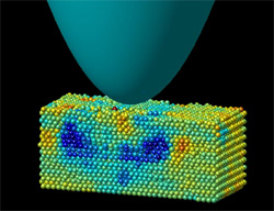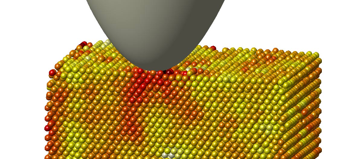
We visualize dislocation nucleation in colloidal crystals in real time and on length scales much larger than the atomic scale : Suspensions of micron-size colloidal particles exhibit Brownian motion and form crystals to increase their entropy. The particles can be tracked precisely in three dimensions using optical microscopy. We grow large single crystals by slowly sedimenting the particles, and we use a spherical indenter (an ordinary sewing needle!) to exert small pressures on these crystals to introduce defects. We can resolve individual thermally driven fluctuation events and see how the applied pressure and thermal fluctuations finally lead to the nucleation of a stable defect (picture on the left).
We also determine the strain distribution from the particle positions in the crystal lattice. This strain is the driving force for dislocation nucleation; these results therefore elucidate the interplay between the applied and local strain and thermal fluctuations that governs the nucleation of these defects. The pictures on the right show how the strain distribution changes when the first defect is formed.

Elastic strain field (shear strain) induced by indenter)

After defect has nucleated:
Strain field of the dislocation loop intersecting the plane.
We also follow the evolution of the defect structure on a larger scale by laser diffraction microscopy. This technique visualizes distortions in the crystal lattice similar to imaging of crystal defects by transmission electron microscopy (TEM) . Although this technique does not allow to resolve the individual particles, it is very useful for obtaining images of the defect configuration alone.

